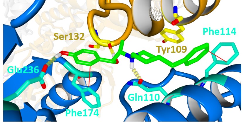
Fig. 4. Ifenprodil (green) re-docked into the X-ray crystal structure of the tetrameric GluN1 (orange) / GluN2B (blue) NMDA receptor (PDB 4PE5). H-bond interactions with Ser132 (yellow) from GluN1 as well as H-bond interactions with GluN2B residues Gln110 (cyan) and Glu236 (cyan) marked by yellow dashed lines. Aromatic interactions with Phe114 (cyan) and Phe176 (cyan) are shown by red lines.